☘️Antinuclear Antibodies...

Highlights
- ☘️Antinuclear Antibodies (ANA)☘️ -Part ✌️
We learnt about the history and lab methods in part ☝️
https://t.co/m7myleJD2I
Let's continue the story!!
#RheumTwitter #MedEd #MedTwitter #rheumatology
(1/24)
 (View Tweet)
- The biggest advantage of indirect immunofluorescence is the ability to gauge antibody specificity from the staining pattern
Prerequisites
🔹An experienced professional
🔹+ & - controls
(2/24) (View Tweet)
- 1️⃣👀 at the uniform staining & staining of the dividing cells(🔺) associated with Ab to histone, nucleosome & dsDNA– characteristic of #lupus 🦋
(3/24)
 (View Tweet)
- 2️⃣Next, a beautiful pattern
Clue: Classic of Limited Systemic Sclerosis
(4/24)
 (View Tweet)
- Any guesses?
(5/24) (View Tweet)
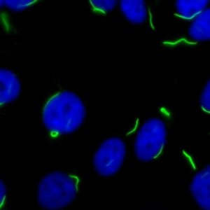
- It's indeed centromere pattern!
Note the discrete speckles (40-80/cell) with intense staining of the metaphase end plate(🔺)
Classic of Limited #Scleroderma
(6/24) (View Tweet)
- 3️⃣More speckling throughout the nucleoplasm 🫥
(7/24)
 (View Tweet)
- What's this pattern called?
(8/24) (View Tweet)
- It's fine speckled!
Fine, numerous speckled throughout the nucleoplasm +/- nucleolar staining
Classic of anti SSA, SSB Ab & some more (Mi2 TIF1y and Ku !)
(9/24) (View Tweet)
- 4️⃣Next🔬
Speckles again but they are coarse this time!
Can you tell me the associated antibodies?
(10/24)
 (View Tweet)
- Select the ✅ combination
(11/24) (View Tweet)
- The correct answer is 3!!
5️⃣Next🔬📷
homogenous?
centromere?
Or just speckled?
This pattern is called 'diffuse fine speckled/DFS' associated with anti-DFS70 Ab
Note the speckled staining of the nucleoplasm
(12/24)
 (View Tweet)
- How is DFS different from FS?
Look at the coarse speckled staining of the metaphase end plate(🔺)!
It can be confused with homogenous pattern too, looking at the 🐀 liver helps!
This Ab is negatively associated with autoimmune disease
🔔🔔🔔 +ANA🚫=autoimmunity
(13/24) (View Tweet)
- 6️⃣Another pretty one!
Which pattern X antibody combination is the correct one??
- Multiple nuc dots a) PmScl
- Coarse speckled b) NXP2
- Nucleolar c) fibrillarin
(14/24)
 (View Tweet)
- Think carefully!
(15/24) (View Tweet)
- It's 1b!
6-20 discrete dots in the nucleoplasm is classic of multiple nuclear dots......
7️⃣ This🔬📷 shows staining of the nucleoli (3-5/ nucleus)
Classic of all these Ab:
🔹Pm/Scl
🔹Th/to
🔹Fibrillarin
🔹RNA pol I
(16/24)
 (View Tweet)
- All the above were nuclear patterns ☢️
We can also have cytoplasmic staining- often in idiopathic inflammatory myopathies especially antisynthetase syndrome ☘️
(17/24)
 (View Tweet)
- We've left out one other important pattern that is a combination pattern 🍹
🔹Speckled nucleoplasm
🔹Metaphase chromosomal staining🔺+ staining of the NOR ⭕
🔹Cytoplasmic staining↗️
🔹+- nucleolar staining
⭐ anti topoisomerase/Scl70 in diffuse systemic sclerosis ⭐
(18/24)

 (View Tweet)
- & then some patterns are just nice to know !!
Tell me which one are these?
(19/24)
 (View Tweet)
- The ✅ combination is:
(20/24) (View Tweet)
- ✅ answer ☝️is 1.
Now, is a positive ANA enough to start thinking of autoimmune diseases?
Or a negative ANA enough to rule them out??
Of course not! 🚫🙅❌
(21/24) (View Tweet)
- Remember, healthy individuals can also have a positive ANA!!
Other autoimmune diseases (as common as hashimoto's thyroiditis, ITP) can also have a positive ANA
#MedTwitter
#MedEd
(22/24)
 (View Tweet)
- Other common causes
Malignancies: most common hematologic like multiple myeloma
Infections: infective endocarditis, chronic– HIV, tuberculosis, Hansen's, acute viral infections
(23/24) (View Tweet)
- Lastly, myositis and most vasculitic syndromes are two important systemic autoimmune diseases which may not have a positive ANA!
Most important bottom line: Clinical correlation is the 🗝️!
Further reading:
https://t.co/umzZxebzSr…
https://t.co/9YnxMUOsZn
(24/24) https://t.co/QuGUyFGKKM (View Tweet)